Thank you to our Funding Sources:
- R01 from the NIH/NIGMS (Miller PI, 9/1/20-8/31/24) “Maintenance of adhesion and barrier function during epithelial cell shape changes”
- Associate Professor Support Fund (Miller PI, 6/1/20-5/31/22) “Investigating how mechanical forces remodel cell-cell junctions in the vertebrate epithelium”
- R01 from the NIH/NIGMS (Miller PI, 8/1/15-8/31/20) “Regulation of localized RhoA activity in dividing epithelial cells”
- Joint NSF/BBSRC Grant (Co-PIs: Ann Miller, Bill Bement, George von Dassow, Andrew Goryachev, 9/1/16-8/31/20) “Bilateral BBSRC-NSF/BIO: Excitocell: A rewired eukaryotic cell model for the analysis and design of cellular morphogenesis”
- MCubed Project (Team Members: Ann Miller, Deb Gumucio, Sundeep Kalantry; 2015-2016) “Survivosome inheritance in pluripotent stem cells”
- K99/R00 Pathway to Independence Award from the NIH/NIGMS (Miller PI, 1/10-8/15) “Regulation of cytokinesis and tumor formation by RhoA”
- MCubed Project (Team Members: Ann Miller, Mary O’Riordan, Christiane Wobus; 2013-2014) “Creating a competent immune competent intestinal layer in vitro”
- Start-up Funds from the University of Michigan to A.L. Miller (2011-2017)
- Biological Sciences Scholars Program to A.L. Miller (2011-2017)
Our Microscopes:
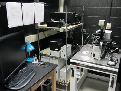 The Olympus Fluoview 1000 Scanning Confocal Microscope is a spectral SIM confocal imaging system on IX81 inverted microscope with 405, 559, and 635nm laser diodes and a multi-line argon laser with a prior motorized XY stage. Our lab’s research relies heavily on high-resolution confocal imaging in 4D (x, y, z, time) or 3D (x, y, time) to image live samples and 3D (x, y, z) to image fixed samples. This Olympus inverted confocal imaging system allows us to image live or fixed Xenopusembryos at multiple stages of development with the speed and sensitivity we need. We often want to image multiple fluorescently-tagged proteins in each cell, and the Olympus spectral detection system allows us to separate multiple fluorophores. In addition, the Olympus SIM scanner technology allows us to efficiently and precisely carry out photobleaching or photoactivation, while simultaneously capturing images immediately pre- and post-stimulation. We can also use the SIM scanner for uncaging caged compounds and for making laser wounds in Xenopus oocytes or embryos.
The Olympus SZX16 Fluorescent Stereoscope has a 16.4:1 zoom ratio with magnifications of 7X to 115X. The 1.0X objective has a numerical aperature of 0.15. It has a LED brightfield light source and a fluorescence light source with an automated shutter for use in time-lapse imaging. The DP72 digital camera can take brightfield color and greyscale fluorescence images with an image resolution of 1.4 million pixels and a live image frame rate of 15 frames per second. This system allows us to view and record still images and live movies of frog embryo cell division, screen for and record images of phenotypes in developing embryos and tadpoles, and identify fluorescent regions in embryos prior to mounting embryos for confocal microscopy.
We also use microscopes at the following University of Michigan core facilities:
Biomedical Research Microscopy Core Facility
Contact: Aaron Taylor (Managing Director) – aata@med.umich.edu
https://brcf.medicine.umich.edu/cores/microscopy/
Biomedical Sciences Research Building (BSRB), Rm A830: 734-763-1170
MCDB Imaging Labs
Contact: Gregg Sobocinski (Manager) – greggps@umich.edu
https://lsa.umich.edu/mcdb/resources/shared-equipment.html
Biological Sciences Building (BSB), Basement imaging suite: 734-615-2034
Our Aquatic Housing Systems:
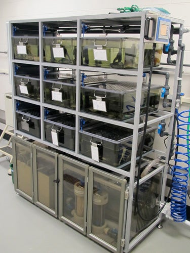 Tecniplast Stand Alone XenopLus Systems house our colony of Xenopus laevis frogs. These recirculating systems maintain consistent, optimal water quality and temperature control so that the frogs housed in the system are healthy. The system draws in well water, filters it, treats it with UV light, and monitors and automatically doses it for pH and conductivity. Automated hourly water changes exchange 10% of the total system water for fresh well water daily. The tanks (and frogs) are easily accessed while on system via a sliding drawer tank system. The tank color (smoky brown) provides an optimal environment for animal health by providing “shelter” and reducing frog stress.
Clones:
Please contact Dr. Ann Miller if you are interested in any of our published clones. We’d be happy to send some DNA to you. Our more popular clones will be made available through Addgene.
Links:
Xenbase is an excellent resource for the Xenopus community which includes information on Xenopus laevis and tropicalis genes and expression patterns, information about Xenopus development, and other useful information for researchers using Xenopus as a model system.
http://www.xenbase.org/common/
The National Xenopus Resource Center (NXR) is located at the Marine Biological Laboratory (MBL) in Woods Hole, Massachusetts, and serves as a national stock center for X. laevis and X. tropicalis as well as a training center for advanced technologies (e.g. husbandry, cell biology, imaging, genetics, transgenesis, genomics).
https://www.mbl.edu/xenopus/
The American Society for Cell Biology (ASCB) brings together cell biologists from all over the country and the world. The ASCB Annual Meeting is an excellent opportunity to hear about the latest cell biology research, network with other cell biologists, and take part in professional development activities. The ASCB also has active committees and provides resources on Women in Cell Biology, Education, Minority Affairs, Public Policy and more.
http://www.ascb.org/
The Cell: An Image Library is a public database of high quality images, videos, and animations of cells. It features cells from a variety of organisms and tissue types. It is a useful resource for cell biology research and education.
http://cellimagelibrary.org/
FIJI is a free image processing package. It brings together ImageJ (free software used in our lab and widely by many researchers for quantification and image processing) with programming capabilities, all the latest plugins, and a user-friendly interface.
https://fiji.sc/
iBiology provides a free and open online platform for communicating the practice of science. The site, which is split into iBioSeminars, iBioMagazine, and iBioEducation sections, holds a growing collection of more than 275 videos by scientists about research and topics related to science. These videos have been viewed more than 3 million times in 170 countries. The project is funded by the National Science Foundation, the National Institute of General Medical Sciences, and the Howard Hughes Medical Institute and is supported by the American Society for Cell Biology and the University of California at San Francisco.
www.ibiology.org