We are developing single-molecule techniques to observe DNA loop formation and breakdown events in vivo. Toward this end, we have devised a novel blend of two-color fluorescence correlation microscopy to observe the behavior of short quantum-dot (QD) end-labeled DNA constructs as they diffuse through a cell. We have been using Dictyostelim Discoideum as a host system, because of the willingness of this aggressive little amoeba to ingest our constructs. The cells are readily imaged under dark-field illumination, while the QD labeled DNA can be seen via fluorescence of the dots after illumination with a 406nm laser.
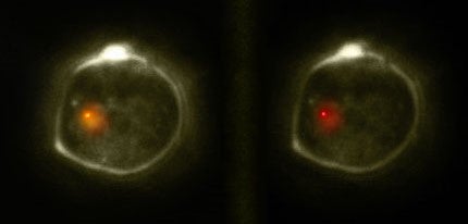
Two-color fluorescence microscopy: Axenic dictyostelium cell transfected with a DNA construct bearing two quantum dots. The simultaneous split-view consists of a 40nm bandpass filter centered at 605nm (left screen) and a 40nm bandpass filter centered at 655nm (right screen)
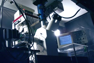
Axiovert inverted microscope customized for dark-field imaging in conjunction with two-color fluorescence.