Live blogger: Ryan Schildcrout
Editor: Brenna Saladin
This piece was written live during the 10th annual RNA Symposium, “RNA Frontiers: From Mechanisms to Medicine” hosted by the University of Michigan’s Center for RNA Biomedicine.

Dr. Nils Walter opens his keynote speech by acknowledging the 10th annual RNA symposium. As a co-founder of the Center for RNA Biomedicine here at the University of Michigan, his excitement for the innovations proposed here is palpable. We feel similarly here at Michigan Science Writers for our 10th year celebration. Walter goes on to say that RNA biomedicine is unique–it offers the fastest path from fundamental discovery to medicine. He emphasizes that this symposium is all about collaboration in working towards swift translation from discovery to medicine.
With the wisdom that science depends on teamwork, Walter emphasizes the importance of building connections between scientists of varied skills, which he says is the key to innovation. He likens scientific collaboration to success on the football field, showing a video of the Michigan football team securing a touchdown, where success in scientific research–and biology itself–depends on the dynamic assembly of its elements.

Turning specifically to RNA biology, Walter challenges the current scientific paradigm of viewing biological structures as stable complexes. For example, cryo electron microscopy (cryo-EM) is able to show the 3D structure of a protein complex, but it does not necessarily capture how that complex may change shape in a dynamic environment, either conformationally or compositionally. He says that this shift towards thinking of biomolecules as being dynamic is important for understanding biological processes. Walter compares thinking of complexes in only their thermodynamically stable result seen in cryo-EM structures is like determining if a wide receiver will score a touchdown based on how the players formation before the ball is snapped–it is difficult to predict the end result without seeing how the players move afterwards.

With the speed of a running back in the open field, Walter dives right in to describing the first of three research areas: riboswitches. Riboswitches are RNA structures that recognize ligands extremely specifically, and undergo conformational changes (changes in molecular shape) that signal if a particular protein should be made or not. Riboswitches are almost exclusively found in bacteria. They enable quick circuit feedback loops which can regulate gene expression (the formation of proteins from underlying genetic signals) for two to four percent of bacterial genes. His lab studies a riboswitch that interacts with manganese to control the speed with which proteins are made. By using green and red fluorescent tags, he can see changes between the open and closed state of the riboswitch in real time and measure its kinetics (which he shows a video of simultaneously). This method is called fluorescence resonance energy transfer (FRET).
FRET operates based on a donor and acceptor fluorophore. The donor by itself will give a fluorescent signal (green). When this fluorophore is in close proximity to the acceptor, it will actually give energy away to the acceptor, which then allows the acceptor to fluoresce (red signal). In this way, they can tag different regions of the molecule (in this case different sections of the RNA) and when these come together they will see that signal change in color. Walter then poses the question: how exactly does manganese specifically alter transcription?
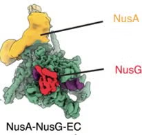
Of course, the kinetics of the riboswitch alone do not tell the whole story. It works in context with transcription, which involves other factors that can impact the riboswitch kinetics. Transcription requires polymerases, which are proteins that facilitate RNA synthesis from a DNA template. He describes a study where they looked at transcription over time with and without manganese, and they found that it is not a perfect “on” or “off” switch. Furthermore, a cofactor NusA, which sits at the exit channel of the polymerase, plays a role in transcription. While cryo-EM experiments show that NusA is stable, Walter found that NusA was constantly binding and dissociating readily. However, within the presence of manganese, NusA doesn’t bind as well. These single-molecule experiments showed the stabilizing effects of manganese and its impact on transcriptional speed via NusA binding. In this way, NusA works to only “tap on the break,” of transcription, if you will, which Walter describes as similar to an anti-lock break system of transcriptional control. The ability of NusA to sense its environment and tap on the break comes from kinetic competition, and to hammer this point home, Walter shows a video of the Michigan football team sacking an Ohio State quarterback.

Walter then talks about a new update to this paradigm, looking at a more complex riboswitch that is sensitive to glycine. Glycine generally stabilizes transcription, but when the binding site is mutated, the switching becomes less prevalent. They performed the same transcription assay where they looked at the products changing over time, and again found that the presence of NusA made the pauses in transcriptional readthrough longer. This suggests that glycine and NusA amplify each other’s activity. Through constant binding and dissociating, NusA displays a uniquely nuanced form of gene regulation. Although the dynamics here are complex, the take-home message is that cooperation between molecules leads to control of transcription, which Walter again uses the video of a Michigan sack against Ohio State to make sure we remember. Walter says that these riboswitches may end up being the “Achilles’ heel” to fight bacteria.
Walter then moves to a different regulatory machine, describing his studies in gene silencing in mammals via miRISC (microRNA-induced silencing complex). While miRNA is known to silence and degrade mRNA, the dynamics surrounding how the miRISC protein complex moves throughout the cell and binds to its targets is still not fully understood. He measured miRISC:mRNA interactions through live cell imaging. Walter found that miRISC performs three-dimensional searching, where it binds, scans a short region to determine if there is a binding site, falls off, and continues repeating this process until it finds a correct binding (in contrast to two-dimensional searching, which would involve binding to mRNA and scanning along its entirety for a binding site, a much slower process). This reversible binding leads to its ability to jump to a large number of mRNAs to regulate, leading to the suppression or degradation of RNA throughout the cell. By looking at the miRNAs bound to the miRISC complex at many time points, Walter proposes that by using the FRET system in this context, he gains an understanding of the kinetics of binding and dissociation, capturing the dynamics of the system. Interrogating miRISC binding transiently could lead to a better understanding of how different regulatory pathways are triggered, such as suppression versus degradation.

The third and final element of the presentation starts with an exploration of the Ship of Theseus in context with biology. The Ship of Theseus begs the question: if all the elements of the ship are replaced, does the ship remain the same? Biology is all about replicating structures using different matter–coding and decoding information that translates to physical action for sustaining life’s essential functions. In this way, there are many different Ships of Theseus in the cell. Walter displays the movie poster for “Everything Everywhere All at Once,” pointing out that everything interacts with each other, and cellular elements that work together over time, or work fast enough, are the interactions that can impact function.
Phase separation, or the compartmentalization of different cellular elements, influences how those elements interact. The Michigan football team appears on the projector again, this time in an on-field brawl, and Walter emphasizes how the dynamics of humans interacting is highly dependent on their proximity to the center of the brawl. Similarly, phase separation in cells has a similar formula, where things stick to each other on the inside of the mass and exchange out on the periphery.

To view this in live cells, Walter presents an experiment where they added high salt to media to create phase-separated particles quickly, which resulted in phase separation for about a third of cellular proteins, an unprecedentedly large number. Walter looked towards analyzing this phase separation by using the FUS protein, which is commonly studied due to its intrinsically disordered region that makes it prone to phase separation. When FUS phase separates, it behaves like a droplet. Interestingly, FUS is often mutated in ALS, which influences its propensity to phase-separate and can contribute to the ALS disease state. When imaging cells with FUS that are phase-separated using salt, they observed that the FUS proteins have tendencies to stay close to certain areas. Proteins and mRNA form into different domains within the droplet, and differences in domain density lead to the molecules being able to travel at different speeds throughout the droplet. These phase-separated droplets can have different textures, and over time, these particles turn from liquid to being gel-like fibers. In doing so, FUS gets stuck in the gel, and can no longer transport to the nucleus to perform its normal functions.

Walter found that these domains, which come from the stickiness stemming from protein folding, are an unintended positive effect of two FDA-approved drugs for treating ALS, where the FUS protein is sequestered to the surface to form fibers, where it will no longer cause disease. He states that this could present a mechanism for therapeutic approaches, where modulating phase separation can help curb diseases that stem from aberrant proteins.
Dr. Walter’s research relies on the idea that biology is dynamic, transient, and cooperative, and he shows throughout his talk that great discoveries will come from appreciating the cell’s ability to control its behavior over time. This idea, which he terms compositional dynamics, is the same driver that leads to success on the football field. As he thanks the auditorium and readies himself for questions, a final video of the Michigan football team scoring a touchdown yields a thunderous applause.
Dr. Nils Walter Bio from RNA symposium website:
Nils G. Walter is the Francis S. Collins Collegiate Professor of Chemistry, Biophysics & Biological Chemistry at the University of Michigan, Ann Arbor, where he co-directs the Center for RNA Biomedicine and directs the Single Molecule Analysis in Real-Time (SMART) Center. Trained at the Technical University of Darmstadt (Diploma with Hans-Günther Gassen) and the Max-Planck Institute in Göttingen (Dr.-Ing. with Nobel laureate Manfred Eigen), he completed postdoctoral work in RNA biophysics before joining the Michigan faculty in 1999 and rising through the ranks.
Walter’s laboratory develops and applies single-molecule and super-resolution fluorescence methods to reveal how noncoding RNAs and RNA:protein machines fold, interact, and function in vitro and in living cells. His group’s methodological and conceptual advances have illuminated heterogeneous folding and catalysis in ribozymes and riboswitches, the biochemistry of the splicing and RNA silencing machineries, intracellular RNA trafficking and RNA:protein complex phase separation, and new paradigms for ultra-sensitive RNA, DNA and protein biomarker detection. These contributions—published in leading journals including Science, Nature, Cell, Molecular Cell, Nature Nanotechnology and Nature Methods—have established single-molecule fluorescence as a central tool for RNA biology.
A committed institutional builder, Walter founded the SMART Center (NSF MRI support) and co-founded the Center for RNA Biomedicine, initiatives that have expanded campus capabilities in RNA therapeutics and advanced microscopy and achieved a national profile. He also served as Faculty Director of the Biomedical Microscopy Core and as Associate Director of the University of Michigan PREP postbac program, and he has a long record of mentoring successful graduate students and postdocs. Walter’s honors include the Camille Dreyfus Teacher-Scholar Award, the RNA Society Mid-Career Award, election as an AAAS Fellow, and institutional awards for mentoring and service. He has received sustained federal support (including an NIH R35 MIRA) and is author of more than 230 peer-reviewed publications (h-index ≈ 69), reflecting a sustained impact on RNA biophysics, translational RNA science, and microscopy.
Ryan is a Ph.D. student in the Biomedical Engineering department at the University of Michigan. His research in Dr. Sriram Chandrasekaran’s lab leverages data-driven machine learning models to study metabolic regulation in cancer. Before starting his doctoral studies, Ryan completed his BFA in music and BSE/MS in biomedical engineering at the University of Michigan. He enjoys running, making music, cooking, hiking, and playing basketball.

To view the original blog post, visit the Michigan Science Writers (MiSciWriters) website at https://misciwriters.com/2026/03/08/dr-nils-walter-life-in-flux-dynamic-rnaprotein-complex-assembly-shapes-biomolecular-function/