Nils Walter and Adrien Chauvier publish new review article – and it’s about time… literally!

Center for RNA Biomedicine Co-Director Nils Walter, Ph.D. is excited about sharing some news about yet another fascinating role RNA plays in gene regulation – and it’s about time… literally. 

In a new review article, “Regulation of bacterial gene expression by non-coding RNA: It is all about time!” published January 10, 2024 in the journal Cell Chemical Biology, Walter and his colleague Adrien Chauvier, Ph.D., at the Single Molecule Analysis Group and Center for RNA Biomedicine, Department of Chemistry, University of Michigan, discuss the vital role that speed plays in the process of non-coding RNA (ncRNA) folding in bacteria.

High-resolution images reveal workings of a bacterial RNA riboswitch, a promising new target for antibiotics

To prevent a global health crisis, scientists around the world are searching for ways to fight bacteria that can evade the current arsenal of antibiotics.

A promising target for new and improved antibiotics are riboswitches, small stretches of RNA that regulate a process necessary for the production of proteins by the bacterial cell. Riboswitches are found almost exclusively in bacteria and could be targeted with antibiotics so that animals or humans are unaffected. With a full understanding of how riboswitches work, researchers may be able to develop drugs that disrupt the cellular machinery that creates needed proteins.

Study: Structural Basis for Control of Bacterial RNA Polymerase Pausing by a Riboswitch and its Ligand

Now, researchers at the University of Michigan’s Department of Chemistry and the Life Sciences Institute have revealed, using a combination of biochemistry, structural biology and computational modeling, how a particular riboswitch regulates its own synthesis.

The first step in generating a protein from the genetic code is called transcription. The enzyme RNA polymerase (or RNAP) travels along the DNA, copying the genetic information found in DNA into a strand of RNA. During this process, RNAP will undergo several “pauses” as it waits for further instructions from the cell. Mechanisms for this pausing and restart have long remained elusive to scientists but promise to become a perfect target for antibiotics.

The team, led by chemistry professor Nils Walter through a collaboration with the labs of LSI professor Melanie Ohi and former U-M scientist Aaron Frank, used a structural biology technique called single particle cryo-electron microscopy (cryo-EM) to visualize for the first time how this transcriptional regulation occurs. Their results are published in Nature Structural & Molecular Biology.

The Walter lab looked at a particular riboswitch that binds a molecule made by the cell, called preQ1. When the preQ1 molecule binds to the riboswitch it alters the shape of the RNA, which then allows the RNAP to once again continue along the DNA so that transcription continues.

Riboswitches were first discovered in 2002, but their specific roles related to the transcription machinery are not well understood. And it’s not hard to see why that is, says Adrien Chauvier, a Walter lab scientist and expert on riboswitches.

“This is a David vs. Goliath situation,” he said. “RNAP is this giant Goliath and the riboswitch is David. Because of this drastic size difference, visualizing where and how preQ1 regulates transcriptional pausing is equal to finding a needle in a haystack.”

Earlier research from the Walter lab revealed that transcriptional pausing is switched on and off as a function of the preQ1 molecule binding to the riboswitch. Moving forward, the Walter lab teamed up with cryo-EM expert Ohi to visualize what was happening.

“This work is a great example of the strength of doing science at the University of Michigan. Three labs with different expertise were able to form a multidisciplinary collaboration that led to an important and novel discovery,” said Ohi, also a professor of cell and developmental biology at the U-M Medical School. “These findings wouldn’t have been possible without this synergy, along with the investments the university has put into strengthening cryo-EM and RNA biology at U-M in recent years.”

Single particle cryo-EM can determine the structures of large protein complexes by building 3D models from millions of 2D images of particles frozen in different orientations, revealing structures that contain molecular details that provide functional insights.

The structural information from single particle cryo-EM corroborated the Walter lab’s earlier findings, but also revealed a specific change in the shape of the riboswitch never seen before. When the preQ1 molecule binds, the riboswitch twists to communicate to the RNAP to continue transcription.

These observations were further rationalized and validated through a collaborative effort with Frank, then a professor of biophysics and chemistry at the University of Michigan and an expert in computational modeling of RNAs. With detailed 3D models in hand, the U-M collaborative team now has a more precise understanding for how this riboswitch regulates transcriptional pausing.

“Now we understand the whole process of riboswitch regulation and can use that knowledge to specifically target these critical parts of bacterial life, hopefully averting the coming crisis of multidrug-resistant bacteria,” Walter said.

In addition to Walter and Chauvier, U-M researchers include Jason Porta, Indrajit Deb, Emily Ellinger and Katarina Mezei. This work was supported by the National Institutes of Health grants to Walter and Ohi, LSI and by the U-M Cryo-EM Biosciences Initiative.

After COVID-19, mRNA vaccines could treat flu, HIV and even cancer

By Justin P. Hicks | jhicks3@mlive.com

Vaccines to protect against severe illness and death from COVID-19 started as the key to a return to normal, but they could wind up unlocking much more for the future of health care.

The mRNA vaccine technology used by Pfizer/BioNTech and Moderna for their respective coronavirus vaccines has been heavily touted by doctors and public health officials as a modern miracle of science and a means to revolutionize vaccine development.

“It’s hard to overestimate the impact this will have on human health,” said Nils Walter, a professor of biological chemistry at the University of Michigan who has studied mRNA for about 30 years. “It’s like introducing the iPhone when everyone had a flip phone.”

Beyond its uses for COVID-19, numerous mRNA vaccine candidates are being researched and undergoing clinical trials for the treatment of cancer, including pancreatic cancer, colorectal cancer and melanoma, as well as HIV, influenza, Ebola, Zika, and rabies.

The same molecule from billions of years ago that gave us life is now coming back to what ails us today in life, which are viral infections and the terrible diseases like cancer,” Walter said. “It’s transformative.”

Vaccines using mRNA technology work by delivering instructions to the cells. In the case of COVID-19, those instructions are to create a protein that mimics the spike protein found in SARS-CoV-2, which triggers an immune response and the development of antibodies to defend against that virus.

Empty syringes wait to be filled with the Pfizer-BioNTech COVID-19 vaccine at the Kalamazoo Expo Center in Kalamazoo, Michigan on Wednesday, May 5, 2021. (Joel Bissell | MLive.com)

Unlike previously used viral vector vaccines, mRNA vaccines don’t introduce live or dead virus into the body as a means of triggering antibody production.

The ingredients of the vaccine are broken down and discarded in a matter of days. The mRNA never enters the nucleus of the cell, and has no interaction with the cell’s DNA, health officials have repeatedly stated.

Dr. Liam Sullivan, an infectious disease specialist for Spectrum Health, expects mRNA technology to “revolutionize vaccine development.”

“This is just the beginning; we’re just scratching the surface here,” he said. “mRNA vaccine technology for treating diseases isn’t going anywhere. If anything, it’s going to get better and better and be used more widespread.”

Previous vaccines like the annual flu shot could take a year to develop, which made it challenging to predict which strains would be around by the next flu cycle. As a result, flu shot effectiveness has varied year to year.

With mRNA vaccines, that timeline can be cutdown 10-fold, Walter said.

“We can now operate at the same speed as the virus and therefore, viruses can no longer outmaneuver us and the vaccine can be made to specs in very, very short time,” he said. “It’s a totally different ball game.”

Continue reading

RNA Society Member Spotlight on Nils G. Walter

“I have a dream. I have been hooked on this dream ever since 30 years ago when I prepared for my German diploma exam by reading a chapter in Lubert Stryer’s Biochemistry textbook about catalytic RNAs” enunciated Dr. Nils G. Walter, a Francis S. Collins Collegiate Professor of Chemistry, Biophysics, and Biological Chemistry at the University of Michigan.

by Dr. Vidhyadhar Nandana

Dr. Nils G. Walter heads the Single Molecule Analysis Group, which integrates cutting edge single molecule microscopy methods, biochemistry, and computational approaches to study dynamics of RNA molecules. Dr. Walter, originally from Germany, obtained his PhD with Nobel Laureate Manfred Eigen in 1995 at the Max Planck Institute for Biophysical Chemistry, Göttingen. He then gained four years of postdoctoral experience studying the biophysics of the hairpin ribozyme with Professor John M. Burke at the University of Vermont before becoming an Assistant Professor and starting his own research group in 1999 at the University of Michigan. He has received numerous awards for his work on RNA, including the RNA Society Mid-Career award in 2017.

RNA has been studied for over 100 years and is connected to the origin of life but “we are still limited in the mechanistic understanding of RNA functions by our incomplete picture of its kinetics,” according to Dr. Walter. He believes that single-molecule tools are perfect to measure these kinetics since they can explicitly observe and measure the folding and unfolding behavior of individual RNA molecules. Owing to his long-standing interest in single-molecule RNA analysis, Dr. Walter has been at the forefront of developing RNA single-molecule tracing techniques in live cells to probe gene regulation. He is the principal scientist in developing single-molecule recognition through equilibrium Poisson sampling (SiMREPS) for the amplification-free digital counting of single unlabeled RNA, DNA and protein biomarker molecules in complex biofluids, which laid the foundation of the biotech startup company, aLight Sciences Inc. Dr. Walter founded and is now directing University of Michigan’s Single Molecule Analysis in Real Time (SMART) Center and the Center for RNA Biomedicine, through which single molecule experiments and RNA research have been accelerated across the university.

Currently Dr. Walter is excited about a research idea that he is hopeful will address both the ongoing pandemic created by SARS-CoV-2 and the” shadow pandemic” of multi-drug resistant bacteria. Many riboswitches pause bacterial RNA polymerase right after they are transcribed, giving the RNA a longer time window to bind its ligand and manipulate the outcome of transcription and translation. Dr. Walter sees an opportunity here to find novel antibacterial agents directed against the RNA polymerase. By applying single molecule and cryo-electron microscopy, he is confident he will also gain insight into the RNA-dependent RNA polymerase of SARS-CoV-2. This work is currently funded by an NIH R35 Maximizing Investigators’ Research Award (MIRA) award.

Dr. Walter took an unusual path into the US academic system. When he started applying for faculty positions, both his PhD and Postdoc mentors were either retiring or no longer fully research active. But the confidence instilled into him by his mentors, including his parents, helped Dr. Walter navigate the process, persevere, and become successful. Immigrating to the USA from Germany and adapting to the new culture was the biggest challenge that Dr. Walter encountered. That being said, he has always been prepared to take on challenges as he feels they build up self-confidence and broaden one’s perspective towards life. He emphasized the greater need for diversity and inclusion in science, which is a fountain of intellectual strength, and this is evident in his own research group’s diversity of people and his service for a decade as his department’s Graduate School Diversity Ally.

“Be bold, be creative, and find your work-life balance to be your personal best. Science is often unpredictable – not to say “frustrating” – but any failed experiment can teach you almost as much as – or even more than – a successful experiment”.

Dr. Walter pointed out the importance of maintaining a balance between one’s professional and personal lives. He has two beautiful kids, and his family is one of his greatest joys. When asked about his good memories with the RNA society, “It was at RNA Society meeting in Banff a friend suggested to me to have kids”, Nils replied with excitement. “The COVID-19 pandemic has further driven home the need for a network of friends to lean on in times of personal or scientific challenges”. Dr. Walter remembers Olke Uhlenbeck’s Lifetime Achievement Award speech in 2006 as his favorite memory at an Annual Meeting of the RNA Society.

“RNA is at an amazing crossroads right now where we finally begin to see its sheer endless power in the form of non-coding, viral, and vaccine RNAs – RNA-seq, siRNAs, as well as CRISPR and RNA medicines are powerful tools”.

Dr. Walter’s favorite RNA is the hairpin ribozyme because he owes it his tenure in the academy. Connect with Dr. Walter on his lab website https://sites.lsa.umich.edu/walter-lab/ and on twitter @NilsWalterLab.

U-M study sheds light on how bacteria control their detoxification

Bacteria need to constantly adapt to compete against other species for nutrient sources and to survive against threats such as antibiotics and toxins.

In an effort to understand how bacteria control and regulate this adaptation, University of Michigan researchers are examining how RNA polymerase—the enzyme that transcribes genetic information from DNA onto RNA—slows during transcription in a process called transcriptional pausing.

They found that a protein called N-utilizing substance A, or NusA, in concert with another control element called a riboswitch, fine-tunes the transcription speed in order to regulate gene expression. Gene expression is the process by which genetic information is converted into the building blocks of the bacterium.

The researchers say their work, published in the Proceedings of the National Academy of Sciences, expands our general understanding of the transcription process in bacteria, and could provide a target for developing new antibiotics.

“NusA is specific to bacteria. It’s not found in human cells or in yeast, so that it can be a target for the design of new antibiotics or drugs that will affect pathogenic bacteria but not our own cells,” said Adrien Chauvier, lead author of the study and postdoctoral researcher in the Nils Walter laboratory in the U-M Department of Chemistry and the Center for RNA Biomedicine.

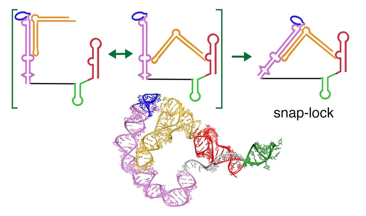
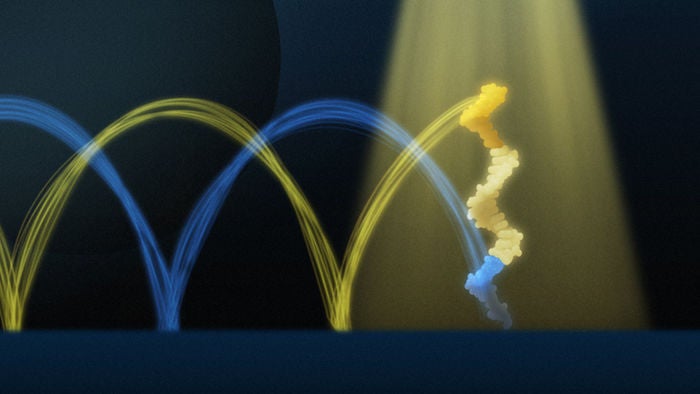
When bacteria undergo gene expression, RNA polymerase synthesizes RNA. As RNA is produced, it undergoes a process called co-transcriptional RNA folding, adopting a dynamic structure that researchers think influences the timing of gene expression. To examine this process, Chauvier, together with undergraduate researcher Pujan Ajmera, looked at an element called a riboswitch, a segment of the transcribed messenger RNA that regulates gene expression through modulation of the RNA conformation. This structural change is triggered when a specific metabolite or ion called a “ligand” binds to the riboswitch.

Chauvier and Ajmera examined a type of riboswitch that binds a fluoride ion and controls the expression of a cellular exporter that removes this toxin from the bacterium. Previously, researchers thought that riboswitches bind to ligands without the influence of proteins, Chauvier said.

“We had the strong idea that riboswitches are also interacting with the entire machinery of the cell, and have the potential to modulate the activity of adjacent proteins to fine-tune gene expression,” he said. “And this is what we found.”

Chauvier, Ajmera and co-authors focused on NusA from the common bacterial species Escherichia coli, because the protein is involved in the three main stages of RNA transcription: initiation, elongation and termination.

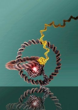
In order to study its role during the transient elongation process, the researchers needed to isolate a transcription complex at a specific position during RNA synthesis. Chauvier developed a method to stop the polymerase during transcription to study the RNA emerging from the RNA polymerase. The researchers then tagged the RNA with a fluorescent marker, allowing individual complexes to be visible under a specialized “single molecule” microscope, and tagged NusA itself.

Additionally, in order to examine the RNA structure, Chauvier and Ajmera used a small DNA probe that transiently binds to the RNA. Using all of these approaches together, the researchers were able to directly watch the frequent, repeated binding of NusA to the transcription complex in real time and correlate the protein’s dynamic behavior to the RNA’s structure. From this, the team discovered that, once the riboswitch binds its fluoride ion ligand, the restructured RNA pauses the RNA polymerase but also suppresses NusA from binding to the enzyme. This speeds up subsequent transcription to more likely express the exporter that detoxifies the bacterium.

“So instead of having a lot of NusA binding events, when the ligand is bound to the riboswitch, we observe less protein binding events, and that turns out to be another way to regulate gene expression,” Chauvier said.

Walter is the senior author of the paper, founding co-director of the Center for RNA Biomedicine, and professor of chemistry, biophysics and biological chemistry.

“There is a tug-of-war between, on the one hand, the fluoride ion binding and folding the riboswitch to then reject NusA protein from the transcription complex and, on the other hand, NusA binding to the complex to suppress fluoride ion binding to the RNA,” Walter said. “Such a dynamic, counteracting modulation of gene expression will allow the bacterium to adapt more nimbly to fluctuating toxin levels. It may also present an expanded angle for developing antibiotics that keep a pathogenic bacterium from detoxing.”

More information:

Read full article >>

PNAS COMMENTARY: The intricate relationship between transcription and translation

Michael W. Webster and View Albert Weixlbaumer
PNAS May 25, 2021 118 (21) e2106284118; https://doi.org/10.1073/pnas.2106284118

Two conserved processes express the genetic information of all organisms. First, DNA is transcribed into a messenger RNA (mRNA) by the multisubunit enzyme RNA polymerase (RNAP). Second, the mRNA directs protein synthesis, when the ribosome translates its nucleotide sequence to amino acids using the genetic code. Because these two processes are so fundamental, a multitude of regulatory processes have evolved to regulate them. Most examples involve regulation of either transcription or translation. In PNAS, Chatterjee et al. (1) instead describe a complex and intricate regulatory process in which transcription and translation are concurrently regulated by each other.

Transcription and translation are commonly viewed as separate. In eukaryotes, their respective confinement to the nucleus and cytoplasm enforces this. Yet, prokaryotes have no such barrier, and newly synthesized mRNAs are translated while they are still being transcribed. RNAP and the trailing ribosome are therefore in close spatial proximity, allowing each to influence the activity of the other. The possibility of a physical connection that could support functional coupling was proposed in 1964 by Marshall Nirenberg’s laboratory based on biochemical experiments (2). They highlighted the potential importance of regulatory processes that simultaneously affect both transcription and translation. Electron micrographs of ruptured Escherichia coli cells, commonly termed “Miller spreads,” confirmed the close proximity between RNAP and the trailing ribosome (3).

Read more >>

Full text in PDF

VIDEO: “The CRISPR Craze: Scientific Breakthroughs Come to the Prepared when Least Expected,” Nils Walter, Ph.D.


The 2020 Nobel Prize of Chemistry recognizes Emmanuelle Charpentier, Max Planck Unit for the Science of Pathogens, Berlin, Germany, and Jennifer Doudna, University of California, Berkeley, USA, “for the development of a method for genome editing.”

Every year, the University of Michigan Complex Systems invites U-M faculty to comment about the Nobel Prizes awards. In this recorded lecture (37 min.), “The CRISPR Craze: Scientific Breakthroughs Come to the Prepared when Least Expected,” Nils Walter, Ph.D., Francis S. Collins Professor of Chemistry, Biophysics and Biological Chemistry, co-director of the University of Michigan Center for RNA Biomedicine, presents the history of the CRISPR discovery.

Starting in 1987 in Japan, CRISPR systems have been observed and studied independently and at times simultaneously by several research groups around the globe (Spain, France, The Netherlands, USA, Sweden, Austria, and Germany). This led the foundation for the 2012 breakthrough by Charpentier and Doudna to harness a CRISPR system (Cas-9) to cleave and modify DNA at specific sites. This genetic editing discovery is currently revolutionizing therapeutics and foundational research, while raising essential ethical questions.

Watch the Lecture


Machine learning expands single-molecule analysis accuracy and accessibility

Adapted from Center for RNA Biomedicine Press Release, November 19, 2020

The observation of single biomolecules in real-time is crucial for our understanding of the cellular biology that is assembled from these molecules, be they DNA, RNA or protein. The recent development of an array of tools and techniques for single-molecule analysis allows studies at an extremely small scale (nanometers, or 10-9 meters) over short periods of time (from a few milliseconds to a second).

Illustration: Deep learning assisted single molecule fluorescence microscopy data analysis

However, until now, most of such observations required tedious and time-consuming manual data processing of thousands of single molecules. A team of University of Michigan (U-M) Department of Chemistry and Department of Physics scientists, spearheaded by graduate students Jieming Li, now Ph.D. in Chemistry, and Leyou Zhang, now Ph.D. in Physics, developed a deep learning algorithm to analyze data emerging from a single-molecule microscope. The results from this collaboration are published in Nature Communications (November 2020).

The key to their automatic data analysis workflow for single molecules (called “AutoSiM”) is to apply a deep learning algorithm, representing a special class of machine learning or artificial intelligence that contains many layers of a neural network, to single molecule fluorescence microscopy data. First, the pattern of the fluorescence signal is recognized to classify the molecule as either relevant or not. Then, the algorithm distinguishes which segment within the fluorescence signal should be included for further data interpretation. For training, the scientists fed the program with a large dataset that was already analyzed by human experts to teach the algorithm to recognize the correct pattern of fluorescence signals, which reflect the behavior of bio-molecules. Once trained on one dataset, it can quickly be adapted to any new dataset by a short “transfer learning”.

There are three main advantages to using AutoSiM: it is faster, significantly cuts down on research time, and reduces costs. “One of my goals is to free up researchers from doing tedious data analysis so they can better focus on their exciting scientific inquiries,” says Li. With the network, data analysis is more consistent than possible across human researchers (due to individual biases), and is free from data entry errors. The concordance between AutoSiM and manual selection is about 90% and is completely reproducible from day to day.

AutoSiM was developed on two datasets from the lab of Nils Walter, U-M Francis S. Collins Collegiate Professor of Chemistry, Biophysics, and Biological Chemistry. One dataset comprised kinetic fingerprints of single molecules termed SiMREPS time traces. The SiMREPS assay distinguishes surface-immobilized mutant DNA biomarkers of disease from wild-type on the basis of distinguishable fluorescence kinetics when a probe interacts with the mutant sequence, wild-type sequence, or surface itself. This molecular diagnostics tool is now being commercialized by aLight Sciences Corp., co-founded by Walter and co-author Alexander Johnson Buck. The other set consisted of time traces characterizing the shape changes of 4 different biomolecular complexes using single molecule FRET (smFRET). Since its development over two decades ago, smFRET has been used to study the dynamics of many biomolecules at the nanometer scale, especially conformational changes involving nucleic acids and/or proteins.

The workflow allows for transfer learning, which means that it can be adapted to new systems by learning from small additional datasets, expanding the original capabilities of the algorithm. “This is like our human brain that learns a lot of new information at a young age, but can also readily adopt new input over its entire lifespan,” says Walter. “It’d be great if others would include their own data to train and use AutoSiM, so the capabilities of the network can be expanded,” explains Li.

The newly released software is available and free for academic purposes, through the U-M Library Deep Blue data repository.

The University of Michigan (U-M) supports state-of-the-art single molecular microscopy at the Single Molecule Analysis in Real-Time (SMART) Center, one of the two core facilities of the U-M Center for RNA Biomedicine. The SMART Center is a shared-use facility providing university researchers with single molecule detection and manipulation tools to track and analyze biomolecules with unprecedented detail. The SMART Center provides access to instrumentation, including single molecule spectroscopy and imaging, laser tweezers, and atomic force microscopy; as well as experienced support in experimental planning and analysis.

Paper cited:
Classification and segmentation of single-molecule fluorescence time traces with deep learning, Jieming Li, Leyou Zhang, Alexander Johnson-Buck and Nils G. Walter, Nature Communications, (2020)11:5833, doi.org/10.1038/s41467-020-19673-1

Methods Editorial: “Introduction to “Convergence of Science and Technology: Fluorescent Resolution of Single RNA Molecules”

As Phillip Sharp, Tyler Jacks and Susan Hockfield of MIT posit in a 2016 report [1]: “The life sciences are in the midst of a revolution… The Convergence Revolution promises to enhance quality of life worldwide … as a result of the sharing of methods and ideas by chemists, physicists, computer scientists, engineers, mathematicians, and life scientists across multiple fields and industries. It is the integration of insights and approaches from historically distinct scientific and technological disciplines.” Perhaps nowhere is this both more apparent and more needed than in taking advantage of the rapid expansion of the RNA biosciences universe and the resulting opportunities for translating discoveries into precision medicines [2]. Converging technological advances, ranging from single-cell RNA sequencing to single-molecule particle tracking at super-resolution, are laying the foundation for reaching an entirely new level of insight into cellular physiology that eluded us just a few years ago [3]. Beyond the emerging catalogues of cellular contents, mechanistic studies of the diverse modes of action of RNAs and their interaction partners are handing us the keys for accelerating progress toward shaping our own destiny. The better we understand the scope and limitations of these new tools, the better we will be equipped to wield them effectively.


Read full editorial in the journal Methods : Convergence of science and technology: fluorescent resolution of single RNA molecules

PDF version

A huge milestone for RNA research at Michigan! U-M Biosciences Initiative invests $45M in ‘groundbreaking’ research (Michigan News)

October 29, 2018

University of Michigan Biosciences Initiative

ANN ARBOR—A new center for the study of concussions, an institute for global change biology, and a facility to advance the new field of cryo-electron tomography are among the University of Michigan projects to be funded in the first round of investments from President Mark Schlissel’s Biosciences Initiative.

Read full article in Michigan News here : U-M Biosciences Initiative invests $45M in ‘groundbreaking’ research

Nature Reviews Research Highlight: “Signals from single molecules”

Mutated DNA strands — even those with only a change in a single nucleobase — can be the cause of serious health problems. Quantifying these mutant DNA strands is particularly challenging when they are present in low concentrations in a patient’s urine or blood samples and must be detected against a large background concentration of wild-type DNA. Now, writing in the Journal of the American Chemical Society, Nils Walter and co-workers report the development of an amplification-free method that they call single-molecule recognition through equilibrium Poisson sampling (SiMREPS).

“We were seeking a powerful tool to detect mutations that are cancer causing, or that make cancer harder to treat,” explains Walter. “One such mutation is found in the epidermal growth factor receptor (EGFR), where a cytosine-to-thymine mutation in the DNA results in substitution of a threonine with a methionine in the protein. The ultimate result of this change is that certain classes of tyrosine kinase inhibitor (TKI) drugs no longer work.”

Read full research highlight in the journal Nature Reviews Materials : Signals from single molecules

PDF version

Nils quoted in Genetic Engineering & Biotechnology news article “Get a Solid Grasp on Tissue RNA Analysis

“Modern techniques like RNA sequencing run into the challenge that low-expressed RNAs are difficult to detect, but often they are more important than the abundant ones,” observes Dr. Walter. “We need tools to look at them.” In a recent study, Dr. Walter and colleagues developed fluorescence-based tools to examine the subcellular location, integrity, and activity of microRNA at single-molecule resolution. “We can localize single microRNA molecules in cells, track them, measure their diffusion constants, and ask how quickly they move between sub-compartments,” asserts Dr. Walter

Full text in pdf format.

Nature Cell Biology “News and Views” : RNA takes over control of DNA break repair

Francesca Storici and Ailone E. Tichon

Small RNAs generated at DNA break sites are implicated in mammalian DNA repair. Now, a study shows that following the formation of DNA double-strand breaks, bidirectional transcription events adjacent to the break generate small RNAs that trigger the DNA damage response by local RNA:RNA interactions.

Maintenance of DNA integrity is crucial for a cell to have a healthy life and for transmission of accurate genetic information to its progeny. Exogenous agents, including radiation or chemicals, as well as endogenous sources, such as reactive oxygen species or defects in DNA metabolism, pose threats to genome stability. Among the most dangerous DNA lesions are DNA double-strand breaks (DSBs), which if not properly and timely sealed can become sites of mutations or chromosomal rearrangements — well-known hallmarks of cancer and other genetic disorders1. The process of DSB repair is one of the most extensively studied mechanisms of DNA repair, yet much remains to be understood about its players and dynamics.
The DNA damage response (DDR) is a complex network of cellular pathways that detect DNA lesions and organize a response signal cascade to repair the DNA. In this issue of Nature Cell Biology, Michelini et al.2 uncover an aspect of the DDR in which RNAs are the directors. The study shows that sequencespecific RNA:RNA interactions orchestrate the DDR in response to DSBs.

Full text in pdf format.

News and Views pice in reference to the Walter Lab NCB paper (ref. 164 at https://sites.lsa.umich.edu/walter-lab/publications)

The drawing shows three different DNA DSB sites in a cell nucleus, depicted as interrupted, thick, parallel strands in blue, green and orange, respectively. The work by Michelini et al.2 suggests that every different DNA DSB site has its unique set of RNA:RNA interactions (shown as small, sandwiched, purple or red rectangles at the two DSB sites on the left) between its specific dilncRNAs and small non-coding RNAs termed DDRNAs (both shown as light blue, green or yellow lines), which originate from dilncRNAs at each DSB site. Such local RNA:RNA interactions are signals to activate the DDR by recruitment of the early DDR factor 53BP1 (transparent ovals). The dilncRNAs generated from DSB ends (thicker lines) were found to be more abundant than those generated towards DSB ends (thinner lines). ASOs can be designed to specifically block DSB repair at a chosen locus, without interfering with DSB repair at other loci within the same nucleus. Here, ASOs (shown as short, thick, brown lines) are specific to dilncRNA sequences of the DSB site on the right and block formation and function of DDRNAs at this DSB site by interacting (small, sandwiched, grey rectangles) with the complementary dilncRNAs.

Nature Reviews Research Highlight: DNA NANOTECHNOLOGY | Head over heels

Dynamic DNA nanotechnology enables the design of DNA-based nanomachines, such as molecular motors or nanorobots. However, most DNA nanomachines operate on a slow timescale, ranging from minutes to hours. Now, Nils Walter and colleagues, writing in Nature Nanotechnology, have made a single-stranded DNA walker that moves by performing cartwheels and at considerably faster speeds than previously reported DNA walkers.

Credit: Rachael Tremlett/Macmillan Publishers Limited

Read full research highlight in the journal Nature Reviews Materials : Head over heels

PDF version


Study finds snap-lock mechanism in bacterial riboswitch

Using single-molecule imaging, scientists from Rice University and the University of Michigan determined that the T-box riboswitch that regulates production of glycine in Bacillus subtilis becomes locked into the “on” position via a snap-lock mechanism. The lock is engaged when an L-shaped arm of uncharged tRNA snaps into position. When the arm is “charged” with a glycine molecule, the arm cannot lock into position, and the switch remains “off.” (Image courtesy of E. Nikonowicz/Rice University)

Finding by Rice, Michigan chemists could point way to new antibiotics
HOUSTON — (May 21, 2018) — In a discovery that points to potential new antibiotic medicines, scientists from Rice University and the University of Michigan have deciphered the workings of a common but little-understood bacterial switch that cuts off protein production before it begins.

Many gram-positive bacteria use T-box riboswitches to regulate production of proteins that utilize amino acids, the basic building blocks of all proteins. A study in Nature Communications describes how one of these switches, a glycine regulator in Bacillus subtilis, flips and locks into the “on” position via a snap-lock mechanism. Engaging the lock increases production of proteins that utilize glycine, the simplest amino acid. Researchers also detailed the switch’s “off” position: A single glycine at the tip of the locking arm blocks protein production.


Read full text here: Study finds snap-lock mechanism in bacterial riboswitch

Nature Research Highlight: Gymnastic feats help DNA ‘walker’ set speed record

A molecular motor flips end over end to cover 300 nanometres a minute.

A short segment of DNA anchors one end of itself and then flings the other end forward. Credit: Zhuoru Li

An acrobatic DNA segment that cartwheels across a surface is one of the fastest ‘walking’ molecules yet designed.

Researchers seeking to design nanorobots have synthesized DNA molecules that move autonomously. But most of these ‘walkers’ require several minutes to take a single step.

Nils Walter at the University of Michigan in Ann Arbor and his colleagues built a DNA walker that moves along a grid of ‘footholds’, which are made of complementary DNA building blocks called nucleotides. Each end of the walker has sequences of nucleotides that it uses to attach itself to the grid. Then it flips end over end to attach itself to another foothold.

The molecule can flip as many as 43 times per minute, covering a distance of 300 nanometres. This is an order of magnitude faster than the pace of other types of DNA walker.

Read full research highlight in the journal Nature : Gymnastic feats help DNA ‘walker’ set speed record

Tiny nanomachine successfully completes test drive

Researchers build a one-wheeled vehicle out of DNA rings

The two rings are linked like a chain and can well be recognized. At the centre there is the T7 RNA Polymerase.
Credit: Copyright Julián Valero

Scientists have used nanostructures to construct a tiny machine that constitutes a rotatory motor and can move in a specific direction. The researchers used circular structures from DNA.

Together with colleagues from the USA, scientists from the University of Bonn and the research institute Caesar in Bonn have used nanostructures to construct a tiny machine that constitutes a rotatory motor and can move in a specific direction. The researchers used circular structures from DNA. The results will now be presented in the journal Nature Nanotechnology.

Read full text here: Tiny nanomachine successfully completes test drive

Built for speed: DNA nanomachines take a (rapid) step forward

When it comes to matching simplicity with staggering creative potential, DNA may hold the prize. Built from an alphabet of just four nucleic acids, DNA provides the floorplan from which all earthly life is constructed.

Through a process known as strand displacement, a tiny walking device composed of DNA moves across a surface in a cartwheeling motion. The new device performed this feat more rapidly than any DNA walker designed to date. Credit: Nature Nanotechnology/Nils Walter

But DNA’s remarkable versatility doesn’t end there. Researchers have managed to coax segments of DNA into performing a host of useful tricks. DNA sequences can form logical circuits for nanoelectronic applications. They have been used to perform sophisticated mathematical computations, like finding the optimal path between multiple cities. And DNA is the basis for a new breed of tiny robots and nanomachines. Measuring thousands of times smaller than a bacterium, such devices can carry out a multitude of tasks.

In new research, Hao Yan of Arizona State University and his colleagues describe an innovative DNA walker, capable of rapidly traversing a prepared track. Rather than slow, tentative steps across a surface, the DNA acrobat cartwheels head over heels, covering ground 10- to 100-fold faster than previous devices.

“It is exciting to see that DNA walkers can increase their speed significantly by optimizing DNA strand length and sequences, the collaborative effort really made this happen,” Yan said.

Yan is the Milton D. Glick Distinguished Professor of Chemistry and Biochemistry at ASU and director of the Biodesign Center for Molecular Design and Biomimetics.

The study was led by Nils G. Walter, Francis S. Collins Collegiate Professor of Chemistry, Biophysics & Biological Chemistry, founding director of the Single Molecule Analysis in Real-Time (SMART) Center and founding co-director of the Center for RNA Biomedicine at the University of Michigan, and his team, along with collaborators from the Wyss Institute, the Dana Farber Cancer Institute and the Department of Biological Chemistry at Harvard (all in Boston, Massachusetts).

“The trick was to make the walker go head over heels, which is so much faster than the hopping used before—just as you would see in a kung fu action movie where the hero speeds up by cartwheeling to catch the villain,” says Walter.

The improvements in speed and locomotion displayed by the new walker should encourage further innovations in the field of DNA nanotechnology.

The group’s findings appear in the advanced online issue of the journal Nature Nanotechnology.

Read full text here: Building Motors to Drive Nanorobots