Partial funding from the National Science Foundation, Award #EAR9911352.
Additional funding from EMAL, the Department of Geological Sciences, the College of Literature, Science and the Arts, and the Office of the Vice President for Research.
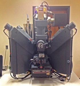
Electron Optics and Imaging
Self-biasing LaB6 cathode.
Two-stage electromagnetic condenser lenses.
Beam regulation from 1-300nA.
Secondary and backscatter electron detectors for electron imaging.
Cameca panchromatic cathodoluminescence detector for CL imaging.
Energy-Dispersive X-ray Spectrometry
Bruker XFlash 2000 silicon-drift energy-dispersive X-ray spectrometer.
Wavelength-Dispersive X-ray Spectrometry
Five wavelength-dispersive X-ray spectrometers with 160mm Rowland circles.
Diffracting crystals capable of analyzing B-U.
Spectrometer 1: LTAP, LPC0
Spectrometer 2: PET, TAP, PC1, PC2.5
Spectrometer 3: LLiF, LPET
Spectrometer 4: LLiF, LPET
Spectrometer 5: LiF, PET, TAP, PC2
Large-area diffracting crystals (spectrometers 1, 3 and 4) for increased sensitivity.
Extended sin(θ) range (spectrometer 5) for increased dynamic range.
Software and Data Processing
ProbeForEPMA, ProbeImage, and Cameca PeakSight software for automated data acquistion and processing.